Detailed guide to eye examination tests including visual acuity, refraction, tonometry, OCT, and more. Know what to expect at Dar El Oyoun Hospital.
Common Eye Tests Explained
Understanding eye tests helps you participate actively in your eye care. Dr. Ahmed Sherif explains each examination component.
Visual Acuity Test
Purpose: Measures sharpness of vision
How it Works:
Read letters on chart (Snellen chart)
One eye at a time
Different sizes of letters
Results noted as 20/20, 20/40, etc.
What Results Mean:
20/20: Normal vision
20/40: See at 20 feet what normal eye sees at 40 feet
20/15: Better than normal
Legal driving limit: Usually 20/40 or better
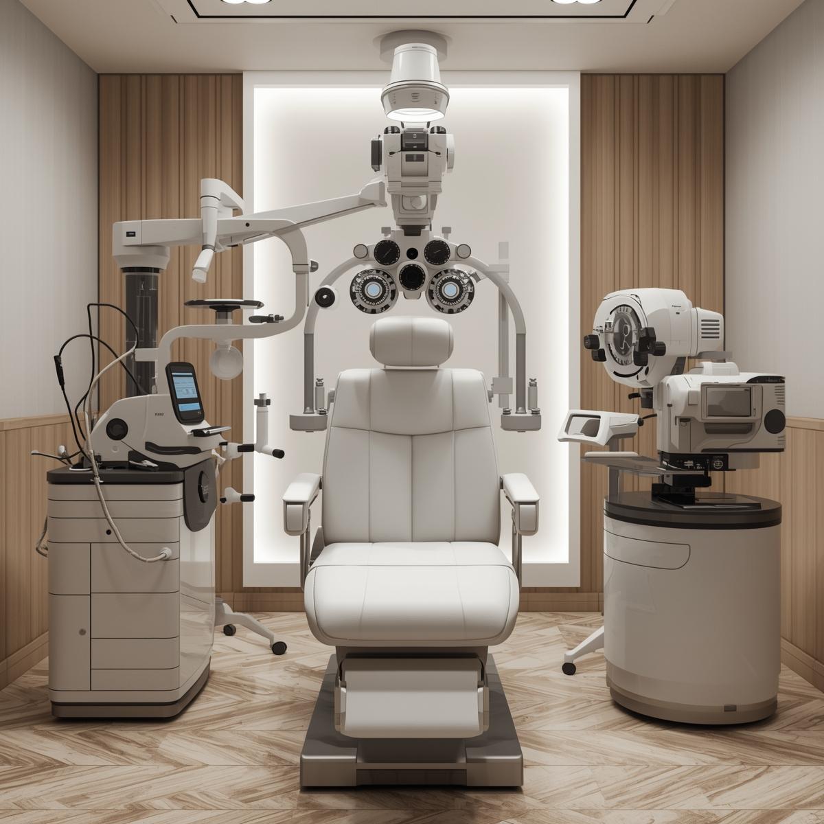
Refraction Assessment
Purpose: Determines eyeglass prescription
Equipment: Phoropter (the big machine with lenses)
Process:
"Which is better, 1 or 2?"
Multiple lens combinations tested
Fine-tunes your prescription
Determines myopia, hyperopia, astigmatism
Output: Prescription with:
Sphere (SPH): Plus or minus number
Cylinder (CYL): Astigmatism correction
Axis: Angle of astigmatism
Add: Reading addition (if needed)
Tonometry (Eye Pressure)
Purpose: Screen for glaucoma
Methods:
Air-Puff Tonometry
Non-contact measurement
Quick puff of air to eye
Mildly startling but painless
Good screening tool
Applanation Tonometry (Gold Standard)
Small probe touches cornea (numbed first)
Most accurate measurement
Used at Dar El Oyoun
Feels like gentle pressure
Normal Range: 10-21 mmHg
High Pressure: Risk for glaucoma
Low Pressure: Can indicate other issues
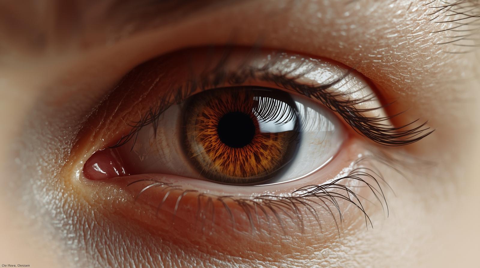
Slit Lamp Examination
Purpose: Detailed view of eye structures
What Doctor Sees:
Eyelids and lashes
Cornea clarity
Iris and pupil
Lens (for cataracts)
Anterior chamber
Tear film quality
Brightlight: Illuminates internal structures
Magnification: Up to 40x zoom
Duration: 5-10 minutes
Comfort: Bright but not painful
Dilated Eye Exam
Purpose: Examine retina and optic nerve
Process:
1. Dilation drops administered
2. Wait 15-30 minutes for full dilation
3. Doctor examines with bright light
4. Detailed retinal viewing
Effects Last: 4-6 hours
Blurred near vision
Light sensitivity
Cannot drive immediately
Bring sunglasses
What's Examined:
Retina health
Macula condition
Optic nerve
Blood vessels
Signs of disease
Optical Coherence Tomography (OCT)
Purpose: Detailed retinal imaging
How it Works:
Like ultrasound but uses light
Cross-sectional view of retina
Measures retinal thickness
Non-invasive, no contact
Detects:
Macular degeneration
Diabetic retinopathy
Glaucoma nerve damage
Macular holes
Retinal swelling
At Dar El Oyoun: State-of-the-art OCT technology
Corneal Topography
Purpose: Maps corneal surface
Uses:
LASIK evaluation
Keratoconus detection
Astigmatism assessment
Contact lens fitting
Post-surgical monitoring
Process:
Look at target in device
Automated scanning
Generates color map
Shows corneal shape irregularities
Results: Detailed elevation and curvature maps
Visual Field Test (Perimetry)
Purpose: Assess peripheral vision
Process:
Look straight ahead at central target
Indicate when you see peripheral lights
Computer maps your visual field
Each eye tested separately
Duration: 5-10 minutes per eye
Detects:
Glaucoma damage
Neurological issues
Retinal problems
Blind spots
Pachymetry
Purpose: Measure corneal thickness
Importance:
LASIK candidacy
Glaucoma risk assessment
Accurate pressure interpretation
Normal Range: 520-580 microns
Quick Test: Non-invasive ultrasound or optical
Color Vision Testing
Purpose: Detect color blindness
Common Test: Ishihara plates
Numbers hidden in colored dots
Different types visible to different color perceptions
Quick screening (1-2 minutes)
Types of Color Blindness:
Red-green (most common)
Blue-yellow (rare)
Complete (very rare)
Autorefractor
Purpose: Quick prescription estimate
How it Works:
Automated measurement
Look at image inside machine
Computer calculates prescription
Starting point for refraction
Advantage: Fast, objective
Limitation: May not be perfectly accurate
Use: Initial assessment, children, verification
Specialized Tests at Dar El Oyoun
Corneal Endothelial Cell Count
Assesses corneal health
Important for cataract surgery planning
Contact lens wear monitoring
Wavefront Aberrometry
Measures optical imperfections
Custom LASIK planning
Higher-order aberrations
Night vision assessment
Fundus Photography
Documents retinal appearance
Tracks changes over time
Diabetic retinopathy monitoring
Patient education tool
Preparing for Your Eye Exam
Before Appointment:
List current medications
Note vision concerns
Bring current glasses/contacts
Know family eye history
Arrange ride if dilation expected
What to Bring:
Insurance information
Previous eye records
List of questions
Sunglasses (for after dilation)
Questions to Ask:
What did tests reveal?
Do I need glasses/new prescription?
Any eye health concerns?
When should I return?
Any restrictions or recommendations?
Frequency Recommendations
Dr. Sherif recommends eye exams:
Age 18-40: Every 2-3 years
Age 40-54: Every 2 years
Age 55-64: Every 1-2 years
Age 65+: Annually
More Frequent If You Have:
Diabetes
High blood pressure
Family history of glaucoma/macular degeneration
Previous eye surgery
High myopia
Taking certain medications
At Dar El Oyoun Hospital
Dr. Ahmed Sherif uses the latest diagnostic technology to provide comprehensive eye examinations with:
Advanced equipment
Detailed assessments
Personalized care
Thorough explanations
Treatment planning
Schedule Your Comprehensive Eye Exam:
Sheikh Zayed: 02-37963013
Dokki: 02-33382136
WhatsApp: +201066479644
Early detection through regular exams protects your vision for life.
Ready for the Next Step?
Book your consultation with Dr. Ahmed Sherif at Dar El Oyoun Hospital